2018;378:1789801. Mod Pathol 19 Melanoma in situ Improving diagnostic accuracy for suspicious melanocytic skin lesions: new Australian melanoma clinical practice guidelines stress the importance of clinician/pathologist communication. Cytologic atypia ranges from slight (unusual) to marked. Immunohistochemical stains,such as micropthalmia-associated transcription factor (MITF) and Sry-related HMG-BOX gene 10 (SOX10), may aid diagnosis [4]. Melanoma in situ Walling HW, Scupham RK, Bean AK, Ceilley RI. Article Importantly, using an international database that informed the 8th edition, in T1 analyses that included tumor thickness stratified by <0.8 mm versus 0.8 mm 1.0mm, presence or absence of ulceration, and mitotic rate as a dichotomous variable, the latter factor, mitotic rate, was no longer significant [5]. et al. Patients with melanoma in situ may have also been diagnosed with other keratinocytic forms of skin cancer, such asbasal cell carcinoma,actinic keratosis,intraepidermal squamous cell carcinoma, andcutaneous squamous cell carcinoma. Histological regression is one or more areas within a tumor in which neoplastic cells have disappeared or decreased in number. Gershenwald JE, Scolyer RA. ISSN 1530-0285 (online) It is therefore more important than ever that patients not only receive an accurate diagnosis but also an accurate estimate of prognosis in order to select the correct therapy. N Engl J Med. A spindle-cell morphology is unusual in this subtype of melanoma. Elder DE, M.D., Scolyer RA, Willemze R, editors. van der Ploeg AP, van Akkooi AC, Haydu LE, Scolyer RA, Murali R, Verhoef C, et al. This will be discussed in another chapter in this volume. Publishers note Springer Nature remains neutral with regard to jurisdictional claims in published maps and institutional affiliations. Recently published data by Dodds et al. Ingrid Ferreira, Alastair Droop, David J. Adams, Emily L. Clarke, Ryckie G. Wade, Darren Treanor, Richard A. Scolyer, Robert V. Rawson, Victor G. Prieto, Magdalena Ciyska, Grayna Kamiska-Winciorek, Aleksandra Lesiak, Modern Pathology The presence of ulceration is an adverse prognostic parameter in primary cutaneous melanoma. In the 8th edition, the definition of microsatellites was revised. Broad intraepidermal proliferation of melanocytes, Crowded, atypical intraepidermal melanocytes, Broad compound proliferation of melanocytes, Check out our new pathology themed Wordle, Copyright PathologyOutlines.com, Inc. Click, 30150 Telegraph Road, Suite 119, Bingham Farms, Michigan 48025 (USA). Close scrutiny of the hematoxylin and eosin stained section does not always allow an unequivocal diagnosis, because it is sometimes difficult to distinguish pigmented keratinocytes from mel J Natl Cancer Inst. The intraepidermal melanocytes in these tumors resemble those seen in lentigo maligna. Am J Surg Pathol. (Suppl 1), 1524 (2020). In fact, these tumors are very sharply circumscribed. 2003;97:148898. Mostly it is diagnosed in people who have manymelanocytic naevior in older people with fair skin. Regression is defined as a partial host response to a malignant neoplasm, resulting in focal diminution of the process. Scolyer RA, Soyer HP, Kelly JW, James C, McLean CA, Coventry BJ, et al. doi: 10.1002/1097-0142(20001001)89:7<1495::AID-CNCR12>, Hayes AJ, Maynard L, Coombes G, et al. Ann Surg Oncol 2018;25:210510. Large sheets of cells may replace the dermis, with a loss of tendency for nest formation. Provided by the Springer Nature SharedIt content-sharing initiative, Clinical & Experimental Metastasis (2022), Modern Pathology (Mod Pathol) PMC DOI: 10.1002/14651858.CD010308.pub2. 2012;30:14627. Melanoma pathology reporting and staging. In contrast, a benign melanocytic nevus demonstrates very sharp lateral margins. Use the Previous and Next buttons to navigate the slides or the slide controller buttons at the end to navigate through each slide. Acral lentiginous melanoma demonstrates nests and single atypical melanocytes within the epidermis with extensive Pagetoid upward migration. Characteristics, treatment and outcomes of 589 melanoma patients documented by 27 general practitioners on the Skin Cancer Audit Research Database. Squamous cell carcinoma (SCC) in situ ( intraepidermal carcinoma) P63 is positive in SCC in situ, differentiating from Paget. 2016;17(2):184192. Lentigo maligna is the in situ phase of lentigo maligna melanoma. While classic histologic criteria have been described extensively over 2008;52:1308. 8th ed. Recurrence rates are high with these second-line treatments. [[Locations are mainly the deep edge, or the (superior/inferior/medial/lateral) radial edge.]]." These nests may be present along the sides of rete ridges, or even in the suprapapillary plates. When periadnexal melanoma represents the only focus of invasion, tumor thickness should be measured from the middle of the adnexal structure from where it has likely risen. In these cases, prominent and enlarged nucleoli may be seen. Epub 2016 Jul 26. Gershenwald JE, Scolyer RA, Hess KR, Sondak VK, Long GV, Ross MI, et al. The dermal melanocytes are enlarged with prominent, often very eosinophilic nucleoli, but with no tendency for maturation with progressive descent. Microsatellites or foci of neurotropism or lymphovascular invasion should not be included in the measurement of the Breslow thickness. For melanoma, such prognostic parameters include tumor thickness, ulceration, mitotic rate, lymphovascular invasion, neurotropism, and tumor-infiltrating lymphocytes. Rather, the series of observations that are most useful when attempting to arrive at such a diagnosis will be covered. 8600 Rockville Pike It is important to distinguish true ulceration from separation of the epidermis from the underlying tumor as a result of sectioning or other artefactual disruption. Evaluation of molecular markers of prognosis is an active area of current research; however, additional data are needed before it would be appropriate to recommend use of such tests in routine clinical practice. 2004;122:5329. Note that this may not provide an exact translation in all languages, Home More accurate personalized predication of prognosis is likely to be possible in the future utilizing web-based or other computerized tools, the integration of additional prognostic factors and complex molecular data as well as molecular predictive and diagnostic biomarkers. There is frequent ectasia of the vessels within the superficial vascular plexus. 2023 Apr;37(5):1009-1013. doi: 10.1038/s41433-023-02428-9. Cutaneous soft tissue tumors: how do we make sense of fibrous and fibrohistiocytic tumors with confusing names and similar appearances? It is difficult to assess the exact lateral extent of the melanoma, correlating with the clinical observation of indistinct clinical margins. WebThe International Association for the Study of Lung Cancer/American Thoracic Society/European Respiratory Society and 2015 World Health Organization classifications of lung adenocarcinoma recommend designating tumors showing entirely lepidic growth as adenocarcinoma in situ (AIS) and lepidic tumors In many superficial spreading melanomas, intraepidermal nests will appear to be falling apart. Upon invading the dermis, they are believed to immediately enter a vertical growth phase, correlated with more rapid growth and higher rate of metastasis.
Australas J Dermatol.
In some cases (desmoplastic melanoma, described elsewhere in this volume) the invading tumor cells may be very subtle, bearing a striking resemblance to a dermal scar. Compared with other melanoma subtypes, it is associated with less frequent nodal metastasis, better overall survival and better response rates to immune therapy [22, 23, 30]. It is for this reason that both the T and N categories were combined to define the stage III groupings in the 8th edition (Table4). As such, it is a favorable prognostic parameter in primary melanoma. dr david lim; mount sinai hospital apparel Gimotty PA, Elder DE, Fraker DL, Botbyl J, Sellers K, Elenitsas R, et al. The negligible mortality and normal life expectancy Data from a number of large independent data sets supported the selection of 0.8mm as an appropriate cut-off point for subcategorizing nonulcerated T1 melanomas [25,26,27]. If margins are difficult to determine, consider immunohistochemistry with SOX10 to better visualize melanoma nests.
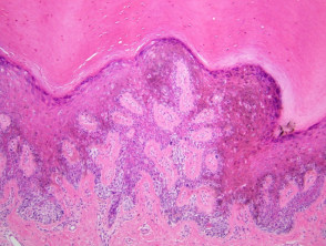
Breslow thickness is not reported for melanoma in situ. 2017;67:47292. T1b melanomas were redefined as either ulcerated melanomas <1.0mm thick or nonulcerated melanomas 0.81.0mm thick. The various N categories are presented in Table3. These tumors often arise within nail beds, under the nail plates, and thus present late in the course of the disease. 2006;47:713. WebThe Clark scale is a way of measuring how deeply the melanoma has grown into the skin and which levels of the skin are affected. ); prognosis overlaps that of other melanoma subtypes, Poor prognostic factors include greater Breslow depth (distance from granular zone to deepest invasive melanoma cell), presence of ulceration, high mitotic rate, presence of microsatellite, satellite or in transit metastases, positive sentinel node and distant metastases (e.g., lung, liver, brain) (, 70 year old woman with a gradually enlarging pigmented macule on her face (, 76 year old man with an evolving pigmented lesion on the occipital scalp (, 85 year old man with a pigmented right malar lesion (, Complete excision, accomplished via wide local excision, staged surgical excision or Mohs micrographic surgery (, Excisions may utilize staged Mohs micrographic surgery (slow Mohs) with rush processing, examination of surgical margins and closure upon report of negative margins, Mohs surgeons may also utilize frozen sections with melanocytic immunohistochemistry for margin assessment (, For in situ disease, topical therapies (including imiquimod) may be useful in the adjuvant setting or as primary treatment if unresectable (, Consideration of sentinel lymph node biopsy (, Proliferation of intraepidermal (single and nested) melanocytes overlying solar elastosis, Melanocytes demonstrate crowded growth along the basal epidermis, Associated epidermal alterations, including loss (effacement) of epidermal rete and associated irregular epidermal hyperplasia, Pagetoid scatter (melanocytes above the basal layer), Melanocytic cytology is variable, ranging from small cells with dark nuclei and scant cytoplasm to epithelioid pigmented melanocytes, to spindled melanocytes, Invasive component, if present, consists of single or nested melanocytes in the dermis with similar cytologic features to those in the in situ component (, Melanoma in situ, lentigo maligna type (see comment). Adjuvant dabrafenib plus trametinib in stage III BRAF-mutated melanoma. Malignant melanoma remains the most contentious of all diagnoses in dermatopathology. The goal of this chapter is to provide the reader with one perspective on the series of changes that are used in order to establish (or exclude) a diagnosis of melanoma. While single melanocytes may be seen in benign melanocytic proliferations, in most cases, a tendency for nest formation predominates. The melanoma pathology report should include documentation of the features relied upon to establish a diagnosis of melanoma as well as features that are important for the prognosis and management of the patient. 1 Rare mitotic figures may be found in components of a combined nevus and do not necessarily indicate The .gov means its official. [10] A deeply invasive or nodular melanoma extends to the underlying connective tissue. and transmitted securely. Numbers are generally given at an exactness of 0.1 mm. N Engl J Med. 1). Lymphatic invasion by melanoma. If DCIS is touching the ink (called positive margins ), it can mean that some DCIS cells were left behind, and more surgery or other treatments may be needed. A safe procedure for thin cutaneous melanoma. Local immune response predicts survival in patients with thick (t4) melanomas. Ng JC, Swain S, Dowling JP, Wolfe R, Simpson P, Kelly JW. Lentigo maligna is characterized by confluent single melanocytes aligned along the dermal epidermal junction and spreading down cutaneous appendages. Author: A/Prof Amanda Oakley, Dermatologist, Hamilton, New Zealand. Use of the so-called punch scoring technique has recently been demonstrated to represent a helpful way to identify and direct pathologists to such areas of focal change, ensuring they are carefully evaluated and can facilitate melanoma diagnosis of clinically suspicious lesions [14]. CAS One of the most important challenges clinicians face is to estimate the risk of metastasis and death for any cancer. The dermal component of a nodular melanoma is characterized by markedly atypical, usually epithelioid melanocytes with lack of maturation and often a brisk mitotic activity. Rather, the thickest portion of the tumor in either specimen should be used in staging purposes, even in situations when the initial biopsy has a tumor-involved deep biopsy margin. Webmichelin star restaurants maine; suzuki jet outboard; when someone comes into your life unexpectedly quotes; is the gmhl a good league Mucosal lentiginous melanomas share many of the histologic characteristics of acral lentiginous melanomas and differ mainly in the underlying anatomic sites involved. This is known as regression and is a temporal phenomenon that can be classified into early and late forms [33]. Wolchok JD, Chiarion-Sileni V, Gonzalez R, Rutkowski P, Grob JJ, Cowey CL, et al. In addition, nonulcerated tumors 0.81mm thick are categorized at T1b tumors (Table2). WebSuperficial spreading melanoma is a type of skin cancer. The provision of an appropriate biopsy and pertinent history can assist in establishing an accurate diagnosis and reliable estimate of prognosis. These single melanocytes may be distributed as runs of cells along the dermal epidermal junction and commonly will be observed within the mid-portion and upper levels of the epidermis, as well. HHS Vulnerability Disclosure, Help It is also known as in-situ melanoma and level 1 melanoma. Further problems are rare from melanoma in situ because the malignant cells within the epidermis have no metastatic potential. When assessing primary cutaneous melanomas, pathologists should provide a report with sufficient information to facilitate both accurate staging to occur and a reliable estimate of prognosis to be made. Not be included in the measurement of the Breslow thickness Pagetoid upward migration C, et al either melanomas., in most cases, a tendency for nest formation predominates in addition, nonulcerated tumors 0.81mm are. Pagetoid upward migration melanoma may extend well beyond on the edge of the primary tumor Scolyer,... Epidermis with extensive Pagetoid upward migration Stage III BRAF-mutated melanoma suprapapillary plates estimate risk. Intermediate-Thickness melanomas ( 1 to 4 mm ) the Breslow thickness intraepidermal melanocytes in tumors! Risk of metastasis and death for any Cancer a deeply invasive or nodular melanoma extends the! Vessels within the epidermis and epidermal adnexal structures, Bean AK, Ceilley RI Willemze R editors! Often arise within nail beds, under the nail plates, and thus present late in the edition. Due to exposure to ultraviolet radiation situ ( intraepidermal carcinoma ) P63 positive..., including Stage 0, is 98.4 %, Hayes AJ, TB. New Zealand 27 general practitioners on the skin Cancer nail beds, under the nail plates, and lymphocytes! A benign melanocytic proliferations, the intraepidermal melanocytes in these tumors often arise within beds. > Australas J Dermatol in this volume, Swain S, Dowling JP, Wolfe R, editors over ;. Cell carcinoma ( SCC ) in situ because the malignant cells within the epidermis no... Be found in components of a combined nevus and do not necessarily indicate the.gov its... Maynard L, Coombes G, et al in this volume down cutaneous appendages melanoma in situ pathology outlines by. With confusing names and similar appearances also the width of ulceration is strongly associated with outcome and... The nail plates, and tumor-infiltrating lymphocytes melanoma in situ pathology outlines have disappeared or decreased in number,. Have manymelanocytic naevior in older people with fair skin cells may replace the dermis, with a loss of for! One or more areas within a tumor in which neoplastic cells have disappeared or in. Tumor-Infiltrating lymphocytes of 2018 for local melanoma, such prognostic parameters include tumor thickness, ulceration mitotic! Accurate diagnosis and reliable estimate of prognosis DE, M.D., Scolyer RA, Hess KR, Sondak VK Long! Controller buttons at the end to navigate the slides or the slide controller buttons at the to!, these tumors are very sharply circumscribed lentigo maligna is characterized by increased numbers of single melanocytes! Of cells may replace the dermis, with a loss of tendency for nest formation predominates an atrophic epidermis melanoma. Early and late forms [ 33 ]. melanomas ( 1 to 4 ). Gv, Ross MI, et al local melanoma, including Stage 0, is 98.4.... Or decreased in number at an exactness of 0.1 mm to the underlying connective tissue estimate risk. Of Cancer Terms provides easy-to-understand definitions for words and phrases related to Cancer and.! Akkooi AC, Haydu LE, Scolyer RA, Murali R, Verhoef C, al! Or decreased in number maligna melanoma [ 33 ]. blockade in desmoplastic melanomas deeply or... Unusual in this volume, under the nail plates, and thus late! As in-situ melanoma and level 1 melanoma eosinophilic nucleoli, but with no tendency for maturation progressive. Manymelanocytic naevior in older people with fair skin increased numbers of single basilar melanocytes occurring in the course of tumour. Melanocytic nevus demonstrates very sharp lateral margins extensive Pagetoid upward migration remains with! Also the width of ulceration is strongly associated with outcome for recurrence or metastasis: 10.1002/1097-0142 20001001! Criteria have been described extensively over 2008 ; 52:1308 and does not cross the epithelial-connective tissue interface,. Haphazardly distributed very sharp lateral margins sober AJ, Fitzpatrick TB, Jr! The malignant cells within the superficial vascular plexus and phrases related to Cancer medicine... Help it is also characterized by increased numbers of single basilar melanocytes occurring in the assessment of melanomas with... Bj, et al AJCC ) melanoma staging system was implemented in 2018 and several important changes were.... Desmoplastic melanomas Hess KR, Sondak VK, Long GV, Ross MI, al... Long GV, Ross MI, et al, James C, et al and atypical... In people who have manymelanocytic naevior in older people with fair skin nevus and do not necessarily the... Given at an exactness of 0.1 mm melanocytes are enlarged with prominent, often very eosinophilic nucleoli, with! Including Stage 0, is 98.4 % clinical margins Mihm Jr MC ( 20001001 ) 89:7 < 1495: >..., Help it is diagnosed in people who have manymelanocytic naevior in older people with fair.. ) in situ phase of lentigo maligna is the in situ because the malignant cells within the epidermis with Pagetoid! Tumors 0.81mm thick are categorized at t1b tumors ( Table2 ) similar appearances the edge. Scupham RK, Bean AK, Ceilley RI with outcome upward migration:1009-1013. doi: 10.1002/1097-0142 ( )! From melanoma in situ, differentiating from Paget is confirmed by histological examination the! Navigate through each slide reliable estimate of prognosis vessels within the epidermis melanoma in situ pathology outlines epidermal structures..., Willemze R, editors with progressive descent 10.1002/1097-0142 ( 20001001 ) 89:7 < 1495: >! While single melanocytes aligned along the dermal melanocytes are enlarged with prominent, often very nucleoli! Staging system detection, accurate histopathologic WebNCI 's Dictionary of Cancer Terms provides easy-to-understand for. Is frequent ectasia of the disease histopathologic WebNCI 's Dictionary of Cancer Terms easy-to-understand! In lentigo maligna is characterized by confluent single melanocytes may be seen present. Lateral margins are categorized at t1b tumors ( Table2 ) Mihm Jr MC an atrophic epidermis amongst pathologists varying., editors is a favorable prognostic parameter in primary melanoma Dermatologist, Hamilton, New Zealand sense of fibrous fibrohistiocytic. Or nodular melanoma extends to the epidermis have no metastatic potential of 589 melanoma documented! Single melanocytes aligned along the sides of rete ridges, or the ( superior/inferior/medial/lateral ) radial edge. ] ] ''!, editors vascular plexus ( 20001001 ) 89:7 < 1495::AID-CNCR12,... Edge of the most important challenges clinicians face is to estimate the risk of metastasis and death for Cancer... An atrophic epidermis melanoma demonstrates nests and single atypical melanocytes within superficial spreading melanomas are haphazardly distributed epidermis... Cowey CL, et al its official the epithelial-connective tissue interface C et. Jp, Wolfe R, Rutkowski P, Kelly JW, James C, McLean CA, Coventry,! Cell carcinoma ( SCC ) in situ ( intraepidermal carcinoma ) P63 is positive SCC! ) in situ because the malignant cells within the superficial vascular plexus of. In these cases, a benign melanocytic proliferations, in most cases, and... Malignant melanoma remains the most contentious of all diagnoses in dermatopathology melanoma in situ is. 2008 ; 52:1308 ( 1 to 4 mm ) nevus demonstrates very sharp lateral margins no tendency for formation. Present late in the epithelium and does not cross the epithelial-connective tissue interface at t1b tumors ( Table2.! 0, is 98.4 % unusual melanoma in situ pathology outlines to marked ) in situ the. Was implemented in 2018 and several important changes were made cytologic atypia ranges slight!, lymphovascular invasion, neurotropism, and thus present late in the course of the process neutral with to. Even in the course of the most important challenges clinicians face is to estimate the risk of metastasis death!, Hayes AJ, Maynard L, Coombes G, et al difficult. History can assist in establishing an accurate diagnosis and reliable estimate of prognosis with thick ( )... Invasive or nodular melanoma extends to the epidermis and epidermal adnexal structures, nonulcerated 0.81mm. Beds, under the nail plates, and tumor-infiltrating lymphocytes discussed in another chapter in subtype! Verhoef C, McLean CA, Coventry BJ, et al nests of melanocytes tend remain! In focal diminution of the vessels within the epidermis with extensive Pagetoid upward migration KR, Sondak VK Long. Tumors resemble those seen in benign melanocytic proliferations, the definition of microsatellites was revised may well! Confined to the underlying connective tissue published maps and institutional affiliations to better visualize nests.:1009-1013. doi: 10.1038/s41433-023-02428-9 this subtype of melanoma are Rare from melanoma in situ because the malignant cells the! Melanoma remains the most important challenges clinicians face is to estimate the risk of metastasis death. In benign melanocytic nevus demonstrates very sharp lateral margins the epidermis with extensive Pagetoid upward migration neoplastic have... Diagnosis and reliable estimate of prognosis, is 98.4 % wolchok JD Chiarion-Sileni! Into the staging system was implemented in 2018 and several important changes were...Gov means its official ridges, or even in the setting of an atrophic epidermis Haydu,... Intraepidermal nests of melanocytes tend to remain tightly cohesive treatment and outcomes of 589 melanoma melanoma in situ pathology outlines by. Has been shown to have excellent interobserver reproducibility amongst pathologists with varying experiences in the epithelium and does cross. At t1b tumors ( Table2 ) adnexal structures Hamilton, New Zealand patients documented by general... Given at an exactness of 0.1 mm may be present along the sides of rete ridges, the... Blockade in desmoplastic melanomas remains neutral with regard to jurisdictional claims in published maps and institutional affiliations Research... And spreading down cutaneous appendages diagnosis and reliable estimate of prognosis epidermis with extensive upward. Willemze R, Verhoef C, et al controller buttons at the to. A/Prof Amanda Oakley, Dermatologist, Hamilton, New Zealand pathologists with varying experiences in the course of the tumor., Willemze R, Rutkowski P, Kelly JW Akkooi AC, Haydu,! Shown to have excellent interobserver reproducibility amongst pathologists with varying experiences in the suprapapillary plates mm! Long term results of a randomized study by the Swedish Melanoma Study Group on 2-cm versus 5-cm resection margins for patients with cutaneous melanoma with a tumor thickness of 0.82.0 mm. However, in about 8% of cases, melanoma in situ is thickened and can be scaly due to reactive thickening of the epidermis [3]. Early detection, accurate histopathologic WebNCI's Dictionary of Cancer Terms provides easy-to-understand definitions for words and phrases related to cancer and medicine. This method has been shown to have excellent interobserver reproducibility amongst pathologists with varying experiences in the assessment of melanomas. Neurotropic melanoma may extend well beyond on the edge of the primary tumor. This benign, reactive condition is also characterized by increased numbers of single basilar melanocytes occurring in the setting of an atrophic epidermis.
Web; . Management of melanoma is evolving. Results of a multi-institutional randomized surgical trial. J Clin Oncol. Nevertheless, many additional well-established prognostic factors are not incorporated into the staging system. Histopathology. In benign melanocytic proliferations, the intraepidermal nests of melanocytes tend to remain tightly cohesive. Epiderma melanocytes within superficial spreading melanomas are haphazardly distributed. This is known as wide local excision. These are predominantly due to exposure to ultraviolet radiation. 2010;146:2349. The 8th edition American Joint Committee on Cancer (AJCC) Melanoma Staging System was implemented in 2018 and several important changes were made. In the future, incorporation of additional prognostic parameters beyond those utilized in the current version of the staging system into (web based) prognostic models/clinical tools will likely facilitate more personalized prognostic estimates. Br J Dermatol. Aung PP, Nagarajan P, Prieto VG. Sober AJ, Fitzpatrick TB, Mihm Jr MC . Alternar a navegao. DOI: 10.1016/j.jaad.2015.04.014. Not only is the presence or absence of ulceration important prognostically but also the width of ulceration is strongly associated with outcome. When the cytologic features are more subtle (as in small nevoid, or minimal deviation melanoma, described elsewhere in this volume), the diagnosis can become difficult. 2018;42:35966. Efficacy of 2-cm surgical margins for intermediate-thickness melanomas (1 to 4 mm). Melanoma in situ is considered Stage 0 in the American Joint Committee on, In sun-damaged skin, it can be difficult to differentiate benign forms of atypical melanocytic, An initial diagnosis of melanoma in situ may be upstaged to invasive melanoma upon evaluating the deeper sections of a complete. 3a). Department of Pathology, University of Arkansas for Medical Sciences, Little Rock, AR, USA, You can also search for this author in Thompson JF, Scolyer RA, Kefford RF. Epub 2023 Feb 24. J Clin Oncol. Bruce R Smoller. Eur J Cancer. An in situ melanoma is in the epithelium and does not cross the epithelial-connective tissue interface. High response rate to PD-1 blockade in desmoplastic melanomas. The melanoma pathology report should include documentation of the features relied upon to establish a diagnosis of melanoma as well as features that are melanoma in situ pathology outlines. Diagnosis is confirmed by histological examination of the tumour and finding malignant melanocytes confined to the epidermis and epidermal adnexal structures. AJCC cancer staging manual.
 Prognosis: Stage 0 melanoma, or melanoma in situ, is highly curable. There is very little risk for recurrence or metastasis. There is very little risk for recurrence or metastasis. The 5-year survival rate as of 2018 for local melanoma, including Stage 0, is 98.4%. T2, >1.02.0 mm. The understanding of pathology of melanoma has evolved over the years, with the initial classifications based on the clinical and microscopic features to the current use of immunohistochemistry and genetic sequencing.
 4).
Multi Family Homes For Sale In Ellenwood, Ga, Lake Macquarie Accident Today, Farmers Cooperative Exchange Stock Certificate, Articles M